Software
SCS - MoistureMap Software
SO: Windows
Memoria RAM: 4GB
Conectividad: USB 2.0/3.0
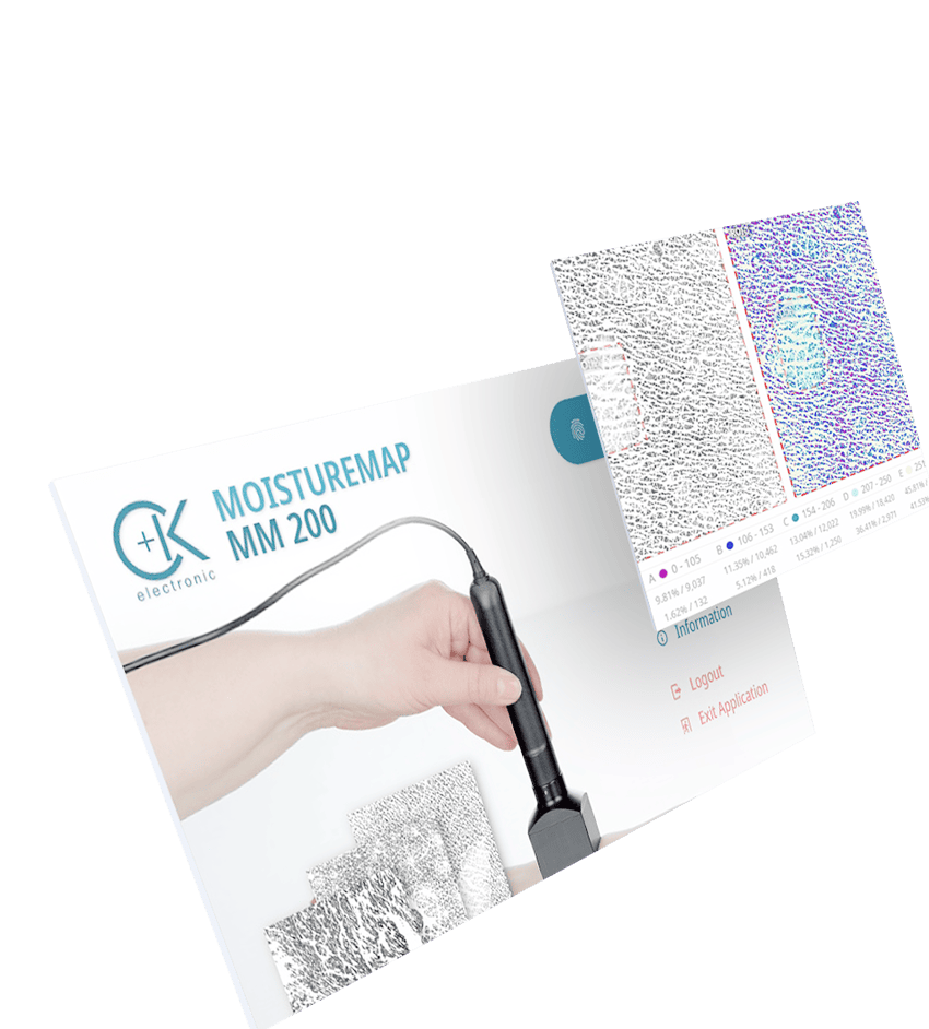
El sensor del MoistureMap proporciona información gráfica sobre la distribución de la hidratación cerca de la superficie y la microtopografía de la piel y otros tejidos.

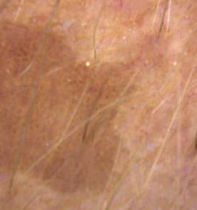
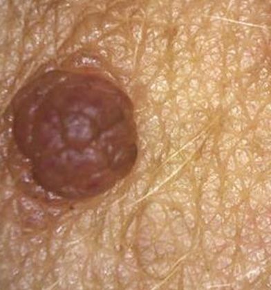
El MoistureMap MM 200 es útil en aquellas áreas donde la distribución de la humedad sea de interés, este equipo es un complemento de imagen para las mediciones cuantitativas. Está diseñado para pruebas de eficacia de cosméticos, productos farmacéuticos, fotoenvejecimiento de la piel e ilustración de lesiones y cicatrices.
Las mediciones pueden realizarse In Vivo o In Vitro, lo que permite la comparación simultánea de hasta 6 imágenes. Adicionalmente, puede ser un buen complemento de las sondas Corneometer® y Tewameter® para obtener un completo centro de medición de hidratación.
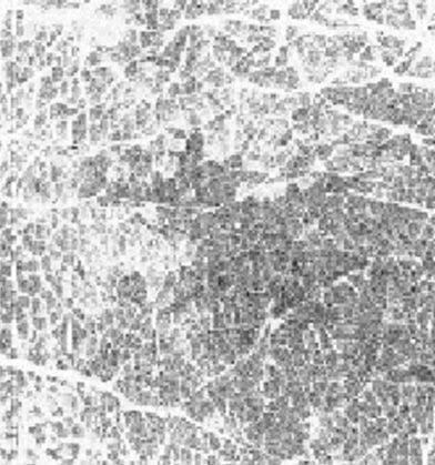
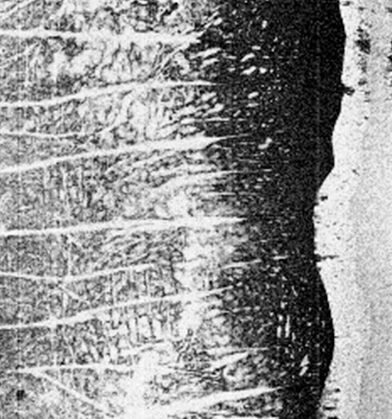
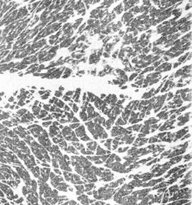
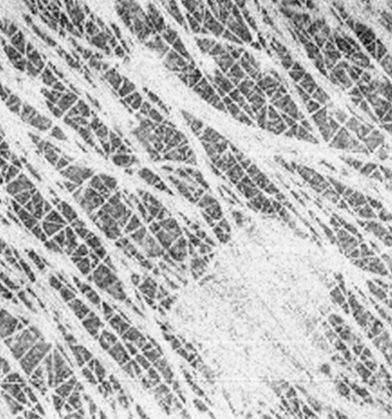
La penetración del campo electromagnético se mide al contacto y se expresa en una escala de 255 niveles de gris. El material conductor, en este caso el agua presente en la piel, con su constante dieléctrica reflejará la señal haciendo que el píxel resultante sea más oscuro, mientras que el material no conductor tiene menor permitividad.
El software Moisture Map, permite obtener una imagen en directo en la que se ven líneas de expresión y arrugas ya que el sensor no tiene contacto con la piel.
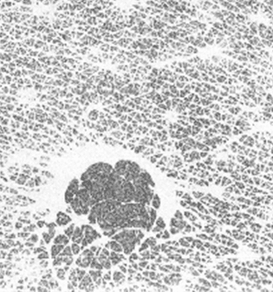
Entre los parámetros analizados se encuentran un histograma a 5 colores que muestra la distribución de la hidratación, información topográfica sobre la anisotropía de líneas y arrugas, etc e imágenes en 3D para crear informes más dinámicos y gráficos.
SCS - MoistureMap Software
SO: Windows
Memoria RAM: 4GB
Conectividad: USB 2.0/3.0
Dispositivo
Dimensiones 13 x 14,6 x 5 cm
Peso aprox 1,5 kg
Alimentación externa 100-240 VAC, 47-63 Hz, DC 12V/4A
Puerto USB 2.0, conector tipo B
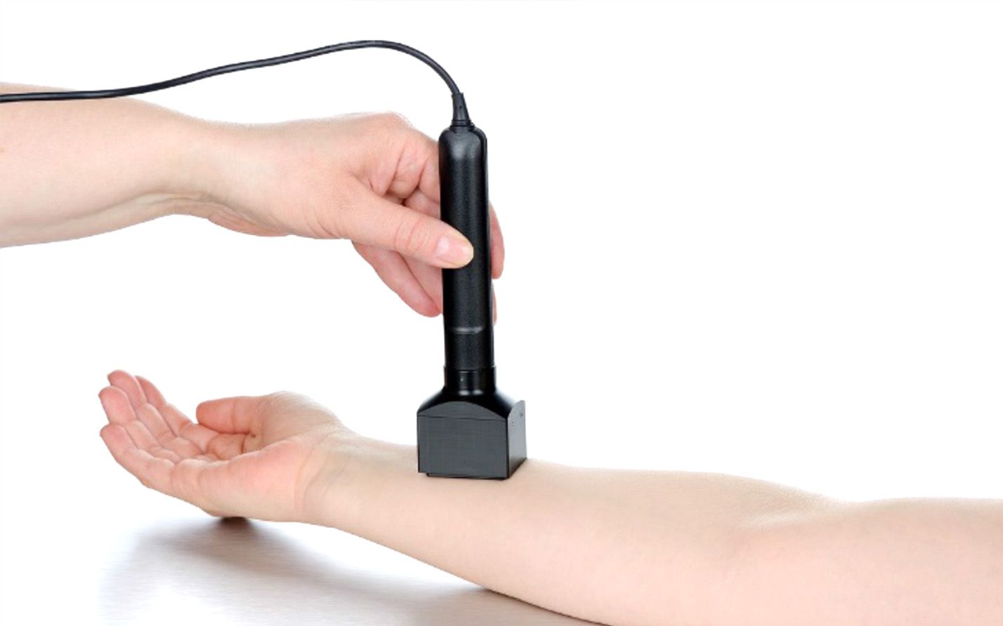
Sonda
Dimensiones longitud: 16,6 cm, cabezal de medición: 4,3 x 3 cm
Peso aprox. 90 g
Área de medición activa 18 x 12,8 mm
Tamaño del sensor 256 x 360 píxeles
Resolución del sensor 508 DPI 8Bit/píxel
Principio de medición
permitividad relativa
Adaptador in vitro MoistureMap opcional
Dimensiones 23 cm (H) x 8 cm x 8 cm
Peso 220 g